Introduction
BM Cleghorn DMD MS
Introduction
On completion of Introduction unit, the student will be able to
1. Define and provide examples of the terms listed below.
Heterodont and Homodont
Monophyodont, Diphyodont and Polyphyodont
2. Describe the different classes and different numbers of teeth in each of the primary and secondary dentitions of man.
3. Describe and identify the bones of the skull that house the dentition.
4. Identify each of the anatomical regions of the mandible and maxilla.
5. Define and describe the "arches".
Maxillary arch
Mandibular arch
6. Understand the numbering of quadrants.
7. Define anterior and posterior teeth.
8. Define and use tooth surface terminology.
|
Vestibular
|
Facial
|
Buccal
|
|
Labial
|
Lingual
|
Palatal
|
9. Define and describe the proper use of proximal tooth surface terminology.
Mesial
Distal
10. Understand and explain the Human Dental Formula.
11. Describe and differentiate the Tooth Tissues.
12. Define crest of curvature (contour) and compare this to contact area.
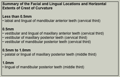
13. Describe the locations and extent of the facial and lingual crests of curvature for the teeth in the secondary dentition.
14. Define and describe the terms line angle and point angle.
15. Define and describe clinical and anatomical crowns.
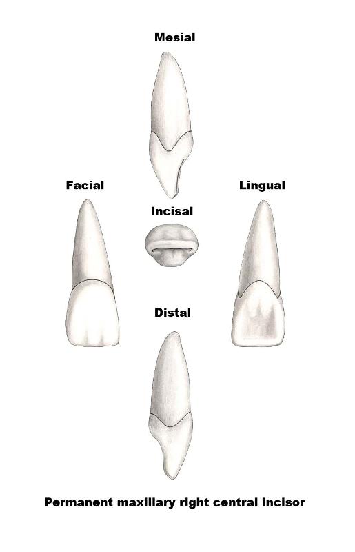
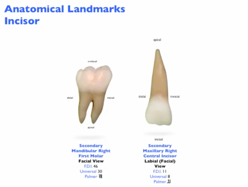
16. Describe the anatomical landmarks of the secondary maxillary central incisor in all views.
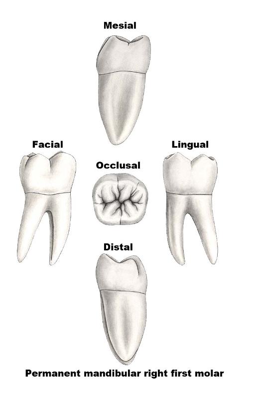
17. Describe the anatomical landmarks of the secondary mandibular first molar in all views.
18. Describe the appropriate order for the naming of teeth (longhand tooth nomenclature).
19. Define and provide examples of
|
Set traits
|
Arch traits
|
|
Class traits
|
Type traits
|
20. Define and describe each of the major shorthand tooth systems and their designations for each tooth in the primary and secondary dentitions.
International (or F.D.I. or Fédération Dentaire Internationale) shorthand system
Universal (American) shorthand system
Palmer/Zsigmondy shorthand system
21. Identify the four basic geometrical shapes of the crowns and summarize the views of teeth with these geometric shapes.
VR Models of the Crowns of the Permanent Teeth (Central Incisors to Second Molars)
VR models of the permanent teeth will be found in each chapter of the Dental Anatomy course. The permanent maxillary right central incisor is illustrated below.
Models for the VR movies provided courtesy of Viade Products, Inc.
Click and hold the cursor down on the 3D model above to rotate the model (or click on the 3D model and use the arrow buttons).
Maxillary right central incisor
Heterodont versus Homodont
Homodont
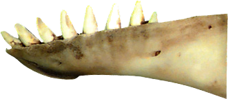 Animals that have teeth that are all the same within the dentition are termed homodonts. An example of a dentition that is classified as homodont is the beluga whale (shown right). All of the teeth of the beluga are anatomically the same. They are conical in shape and interdigitate with each other. Dolphins and sharks are other examples of a homodont dentition.
Beluga whale mandible (sagittal view)
Heterodont
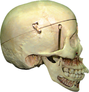
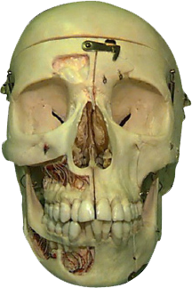
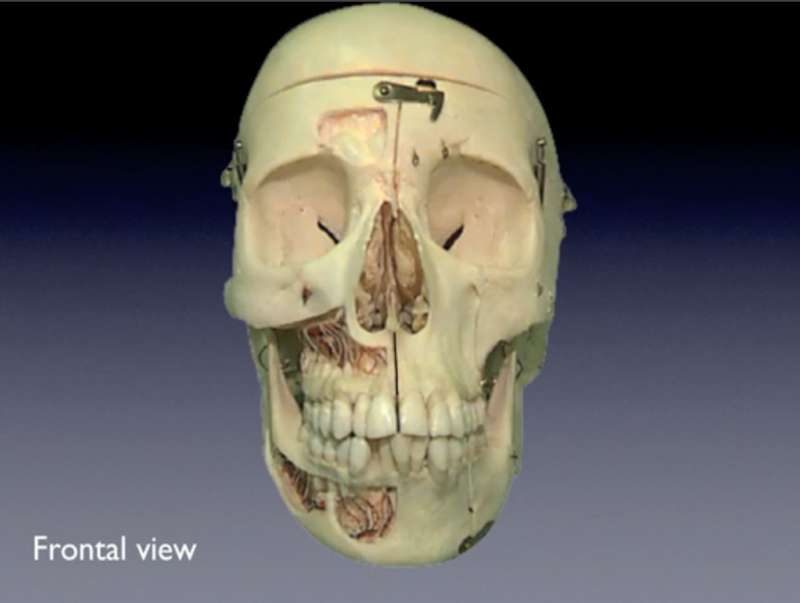
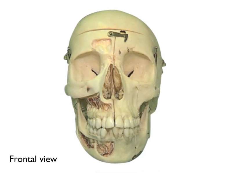
Most mammals including man, however, have teeth that are of different types or classes. The permanent dentition of man is composed of four different classes of teeth (incisors, canines, premolars and molars). Man can therefore be classified as heterodont. The primary (deciduous) dentition of man has only three classes of teeth. Man can therefore be classified as heterodont.
|
|
|
|
Human skull (sagittal view)
|
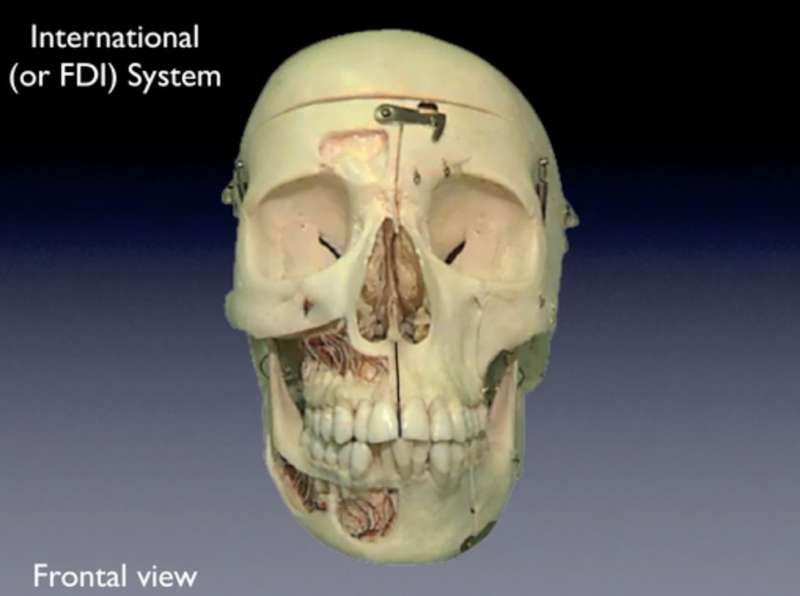
Human skull (frontal view)
|
Monophyodont, Diphyodont and Polyphyodont
Monophyodont
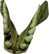 Dentitions can be classified by the number of sets of teeth. Monophyodont means "having one set of teeth". Animals like the beluga whale (shown right) have only one set of teeth in their lifetime. The aquatic mammals (Cetacea) include a group of toothed whales (Odontoceti) that are mostly homodont and monophyodont. Others in this group include the dolphin, the porpoise and the narwhal.
Beluga whale mandible (frontal view)
Diphyodont
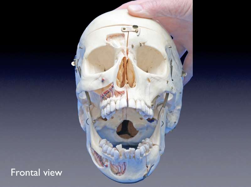
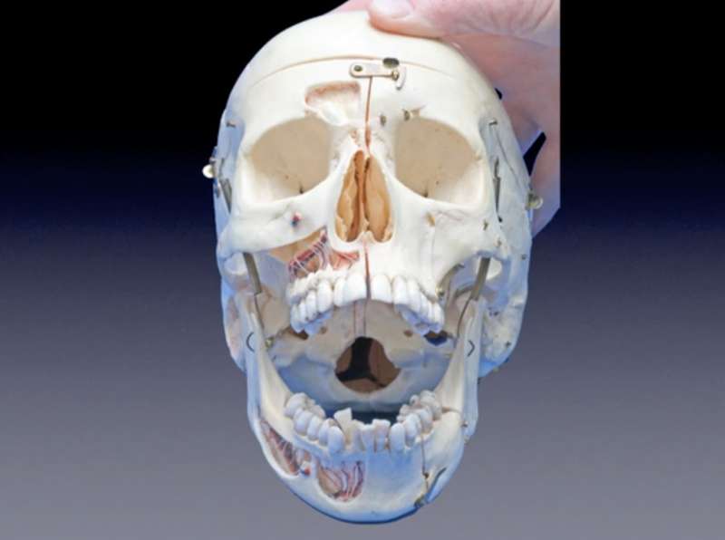
Diphyodont means "having two sets of teeth". Mammals usually have two dentitions and these are termed the primary (deciduous) dentition and the secondary (permanent) dentition. Man is an example of a mammal that is classified as diphyodont. Man has a primary (deciduous) dentition that generally begins to erupt at approximately six months of age. By approximately two and a half years of age, the primary dentition is complete. A total of twenty teeth comprise this primary dentition and are erupted by that time. The secondary (permanent) dentition begins to erupt at approximately six years of age. Both primary and secondary (permanent) teeth are present through a mixed dentition period which lasts from age six to approximately eleven and a half years of age. Secondary (permanent) teeth continue to erupt until the late teens or early twenties. The total number of teeth in the secondary (permanent) dentition of man is thirty-two.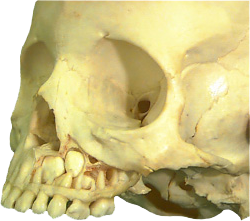 The skull of a 5 year old (shown right) illustrates an intact fully erupted primary (deciduous) dentition in the maxillary arch. Some of the tooth buds of the developing secondary (permanent) dentition are visible.
Skull (5 year old)
Polyphyodont
The term polyphyodont refers to dentitions that have an endless succession of teeth. The dentition of the shark represents the polyphyodont dentition. Teeth are exfoliated or lost and are soon followed by replacement tooth. Amphibians with teeth, such as the frog (Rana temporaria) and the mud puppy (Necturus maculatus) are examples of dentitions that are homodont (teeth all the same) and polyphyodont (multiple sets of teeth). Reptiles are also generally homodont and polyphyodont. The iguana (Iguana iguana) is an example of this type of dentition.
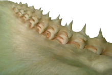
Shark mandible (medial view)
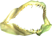
Shark jaw (frontal view)
Maxilla & Mandible
Maxilla
The bones of the skull that provide anchorage for the teeth are the paired maxillary bones and the mandible. The maxillary bones are sutured at the midline and initially house the 10 primary (deciduous) teeth. When the permanent (secondary) dentition is fully erupted, 16 permanent teeth are present in the maxillary arch.

Click on the movie icon above to view the osteology of the skull.
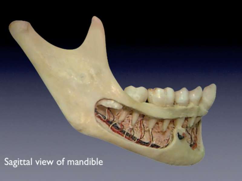
Mandible
The mandible is comprised of specific anatomical regions. The body of the mandible houses the teeth of the mandibular arch. The single u-shaped mandible contains 10 primary (deciduous) teeth and then the 16 teeth of the permanent dentition. The bone surrounding the teeth is called alveolar bone. Loss of teeth results in loss of alveolar bone. The ramus of the mandible is connected to the body and is the region that has the mandibular foramen and lingula on its medial aspect. This is the location for needle placement to anaesthetize the mandibular teeth in a typical mandibular block. The angle of the mandible is the region where the body and ramus meet. The condyle is the bony portion of the complex temporomandibular joint. The u-shaped mandible has two condyles in a ginglymoarthrodial type of joint. The coronoid process provides an attachment for the insertion of the temporalis muscle and the deep masseter muscle. The mandibular notch is the concave area of the superior portion of the mandible between the condyle and coronoid process.
Click on the movie icon above to view the anatomical regions of the mandible.
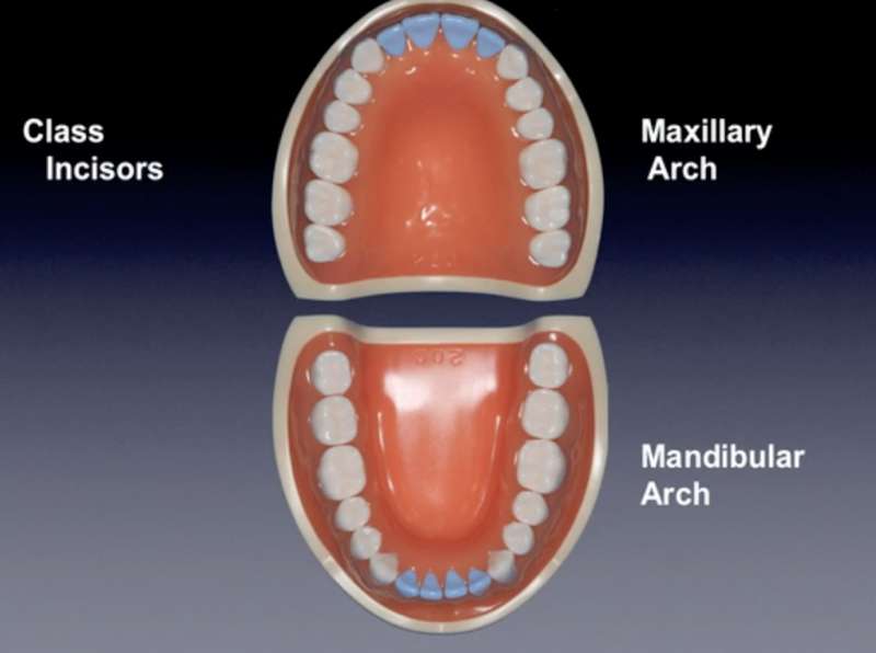
Maxillary and Mandibular Arches
The teeth are housed in the alveolar bone in the respective maxillary and mandibular arches.
Click on the movie icon above to view the arches.
Quadrants
The arches can be divided into quadrants. Each arch contains two quadrants. Quadrants are numbered from 1 to 4. The numbering begins with the maxillary right quadrant and proceeds in a clockwise direction.
Click on the movie icon above to view the quadrants.
Classes of Teeth in the Permanent Dentition
The secondary (permanent) dentition has 4 classes of teeth. They are incisors, canines (cuspids), premolars (bicuspids) and molars.
The class of incisors includes 8 teeth. They are the maxillary left and right central and lateral incisors and the mandibular left and right central and lateral incisors. The class of canines totals 4 teeth, one in each quadrant. They are the maxillary left and right canines and the mandibular left and right canines. The premolars total 8 teeth. Each quadrant has a first and second premolar. There are three molars in each quadrant for a total of 12 molars in the permanent dentition. The permanent dentition, therefore has 32 teeth in total.
Click on the movie icon above to view the classes of teeth in the permanent dentition.
Classes of Teeth in the Primary Dentition
The primary (deciduous) dentition has only 3 of these classes of teeth. These are the incisors, canines and molars. There are no premolars in the primary dentition. The primary dentition will be presented in a later chapter.
Anterior versus Posterior Teeth
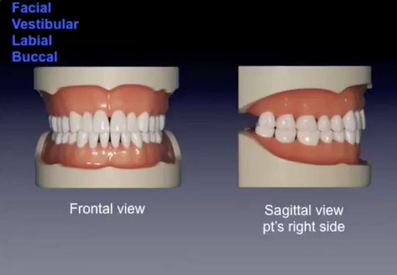
Teeth can also be classified as being either an anterior or a posterior tooth. Anterior teeth include the incisors and canines. The outside surface of the anterior teeth is adjacent to the lips. Therefore the outer surface of the anterior teeth is called the labial surface. Posterior teeth include the premolars and molars. The outer surface of the posterior teeth is adjacent to the cheeks. Therefore the outer surface of the posterior teeth is called the buccal surface.
A more detailed description of tooth surfaces follows this section on anterior and posterior teeth.
Click on the movie icon above to view anterior versus posterior teeth.
The primary dentition includes only three of the four classes of teeth - incisors, canines and molars. Therefore the anterior teeth in the primary dentition include the incisors and canines. The posterior teeth in the primary dentition only include molars as there are no premolars in the primary dentition.
Tooth Surface Terminology
The crown of each tooth has five tooth surfaces. However, there are more than five terms that can be used. Some terms apply to specific surfaces of particular groups of teeth while other terms are more collective. The five views of an anterior tooth (maxillary incisor) and a posterior tooth (mandibular molar) are shown below.
These examples illustrate one of the differences in terminology between anterior and posterior teeth. The "biting" surface of an anterior tooth is the "incisal" surface while the biting surface of a posterior tooth is called the "occlusal" surface. A more detailed explanation of the terms follows.
|
Vestibular or Facial
Buccal
Labial
Palatal
Lingual
Mesial
Distal
|
Click on the movie icon above to view tooth surfaces terminology.
|
To describe specific locations on teeth, and in particular, the crowns of teeth, line angles, point angles and crown thirds are used. These are descriptive terms as teeth are not flat but generally convex or concave surfaces that join together seamlessly
Line Angles
A line angle is the area where two tooth surfaces join together. In the purest sense, a line angle is produced by two flat surfaces meeting. Teeth have curved surfaces using line angles helps in defining specific locations on the crown and root. Line angles are named by the joining of the two surfaces.
Point Angle
A point angle is the junction of three tooth surfaces and is named by the combination of the surfaces involved.

Click on the movie icon above to view laine and point angles.
Crown thirds
All crowns can be divided into a cervical third and a middle third. All posterior teeth (premolars and molars) have an occlusal third while all anterior teeth (incisors and canines) have an incisal third.
Click on the movie icon above to view crown thirds.
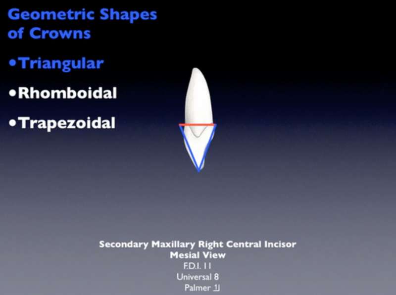
Geometrical Shapes of Crowns
Understanding the basic geometric shapes of the crowns of teeth provides an overview to the more detailed anatomical features of individual teeth. Locations and extent of crests of curvature (contour) of crowns, positions of cusps and incisal ridges will be better appreciated once the geometric shapes of the crowns in each view are understood.
All of the crown surfaces (except for the incisal and occlusal views) can be described using one of three basic geometrical shapes. They are triangular, trapezoidal and rhomboidal.
Click on the movie icon above to view geometrical shapes of crowns.
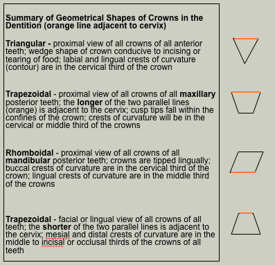
The shapes of crowns from the incisal (anterior teeth) and occlusal (posterior teeth) views will be addressed in the detailed chapters on each individual tooth in the primary and secondary dentitions.
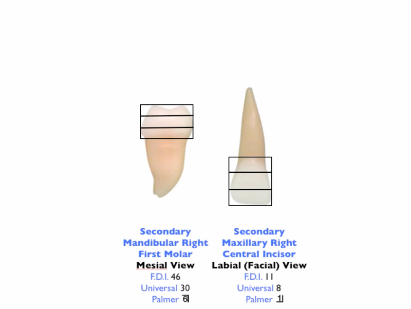
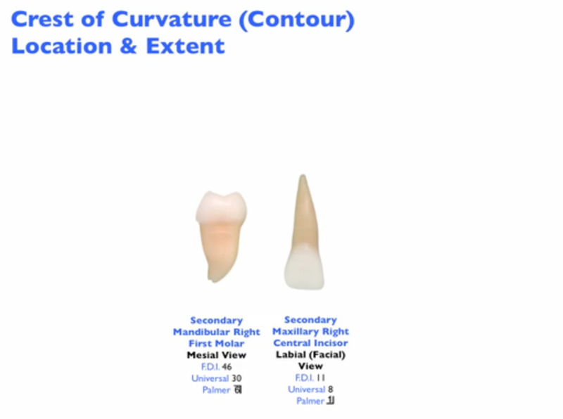
Crest of Contour (Curvature)
Location of the Crest of Curvature
The crest of curvature (also called the crest of contour) is defined as the widest area of the crown of the tooth when viewed from the facial (lingual) or proximal (mesial or distal). Each tooth has a distinct location for the crest of curvature and it is based on the crown divided into thirds (ie. cervical, middle, incisal or occlusal thirds). The crest of curvature is synonymous with the contact area only when teeth are in perfect alignment. When teeth are not in perfect alignment, the contact area will not correspond to the crest of curvature.
Extent of the Crest of Curvature
The extent of the crest of curvature describes how bulbous a tooth is on either its facial or lingual surface. The teeth with the least amount are the mandibular incisors. The greatest extent of the crests of curvature are found on the lingual surfaces of the secondary mandibular molars. The locations and extents of the crest of curvature for each tooth will be described in detail in subsequent chapters.
click on the movie icon above to view extent of crests of curvatures.
Anatomical Landmarks of Teeth
All teeth have a crown and a root. Some of the major anatomical features of anterior and posterior teeth are illustrated below. A more detailed description of the anatomical features of each tooth in the secondary and primary dentitions will follow in subsequent chapters.
Key Anatomical Landmarks
|
anatomical crown
|
mamelon
|
fossa
|
|
anatomical root
|
cingulum
|
pit
|
|
enamel
|
marginal ridge
|
furcation
|
|
dentin
|
lingual fossa
|
cusp
|
|
cementum
|
cusp tip
|
cusp ridge
|
|
cervical line
|
triangular ridge
|
developmental groove
|
|
apex
|
apical foramen
|
incisal ridge
|
|
incisal edge
|
supplemental groove
|
|
Click on the movie icon above to view anatomical landmarks.
Clinical Crown versus Anatomical Crown
The anatomical crown is defined as that portion of the tooth that is covered by enamel. The clinical crown is that portion of the tooth that is present intraorally. There are therefore clinical scenarios where the anatomical crown is shorter than, equal to or longer than the clinical crown.
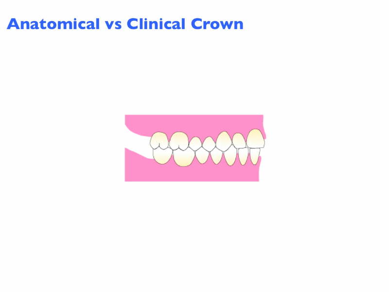
Click on the movie icon above to view anatomical versus clinical crown.
Tooth Numbering Systems
Teeth can be named using either the longhand designation or using one of a number of shorthand systems. When using the longhand designation, teeth should be identified using proper terminology in a precise order. Two examples are shown below.
"Secondary maxillary right first molar"
"Primary mandibular left second molar"
These two examples are indicative of a systematic order of terms from Set (primary or secondary) to Arch (maxillary or mandibular) to Side (left or right) to Tooth (central incisor, first molar, second premolar, etc.).
Using the longhand designation each time a tooth is described would be cumbersome and time-consuming. As a result, a number of different shorthand numbering systems (or designations) have been developed to provide efficient, clear and easy methods to describe each of the teeth in the dentitions. The three most commonly used shorthand systems are described below.
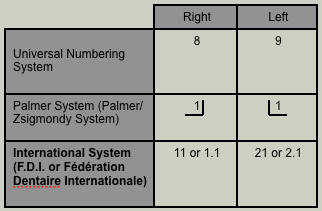
The three shorthand systems that will be described are the International system, the Universal system and the Palmer or Palmer/Zsigmondy system. The table below illustrates the three shorthand designations for the secondary maxillary left and right central incisors.
Some differences that should be noted between the primary and secondary dentitions are as follows:
1. There are 20 teeth in the primary dentition as opposed to 32 teeth in the secondary dentition. 2. The primary dentition has 2 molars per quadrant while the secondary dentition has 3 molars per quadrant. 3. There are no premolars in the primary dentition. The secondary dentition has 2 premolars per quadrant (for a total or 8).
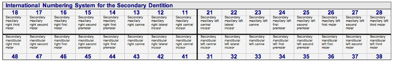
International (F.D.I) Shorthand Numbering System
International (F.D.I) Shorthand Numbering System for the Secondary Dentition
The International (or F.D.I. or Fédération Dentaire Internationale) shorthand system is used throughout the world as the shorthand method of choice except in the United States. This system has two numbers for each tooth. The first number represents the quadrant. Quadrants are numbered in a clockwise manner from the maxillary right to the mandibular right. The quadrants for the secondary dentition in this system are 1 (maxillary right), 2 (maxillary left), 3 (mandibular left) and 4 (mandibular right).
|
FDI System Naming of Quadrants for the Secondary Dentition
|
|
1 = Maxillary right
|
2 = Maxillary left
|
|
4 = Mandibular right
|
3 = Mandibular left
|
The second number in this system represents the secondary tooth from 1 (central incisor) to 8 (third molar).
|
FDI System Naming of Teeth for the Secondary Dentition
|
|
1 = central incisor
2 = lateral incisor
3 = canine
4 = first premolar
5 = second premolar
6 = first molar
7 = second molar
8 = third molar
|
The International system may or may not have a "period" between the quadrant and the tooth. The National Dental Examining Board of Canada includes a "period" between the quadrant number and the tooth number as this allows a clear differentiation between the Universal and International systems. The "official" F.D.I. numbering system, however, does not use a period between the two numbers. Unfortunately, this can sometimes lead to confusion as tooth 24 in the International system is the secondary maxillary left first premolar while in the Universal system tooth 24 is the secondary mandibular left central incisor.
The pronunciation also differs between the Universal and the International systems. The secondary maxillary left first molar in the Universal system is tooth 14 and is pronounced as "fourteen". The shorthand designation for the same tooth in the International system is tooth 2.6 or 26. However, it is pronounced as tooth " two six" in this system.
Click on the movie icon above to view the FDI shorthand system.
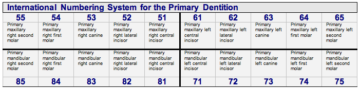
International (F.D.I.) Shorthand Numbering System for the Primary Dentition
The first of the two numbers used in the International shorthand numbering system for the primary dentition is the quadrant number. The quadrants are numbered 5-8 when using the International system for the primary dentition.
|
FDI System Naming of Quadrants for the Primary Dentition
|
|
5 = Maxillary right
|
6 = Maxillary left
|
|
8 = Mandibular right
|
7 = Mandibular left
|
The second number in the International system is the specific tooth. In the primary dentition each quadrant contains a total of 5 teeth. Therefore there are a total of 20 teeth in the primary dentition.
|
FDI System Naming of Teeth for the Primary Dentition
|
|
1 = central incisor
2 = lateral incisor
3 = canine
4 = first molar
5 = second molar
|
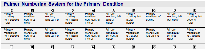
Palmer Shorthand Numbering System
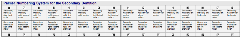
Palmer Shorthand Numbering System for the Secondary Dentition
The Palmer or Palmer/Zsigmondy numbering system uses one of 4 quadrant symbol designations for each of the four quadrants.
|
Palmer System Quadrants
|
|
 = Maxillary right = Maxillary right
|
 = Maxillary left = Maxillary left
|
|
 = Mandibular right = Mandibular right
|
 = Mandibular left = Mandibular left
|
The quadrant symbols are used for both the primary and secondary dentitions.
Each quadrant in the secondary (permanent) dentition has eight teeth. The teeth are numbered from 1-8 in each quadrant with 1 representing the central incisor and 8 representing the third molar. The tooth number is placed within the appropriate quadrant symbol.
For example, the Palmer shorthand designation for the secondary mandibular left first molar is  .
.
Palmer Shorthand Numbering System for the Primary Dentition
The Palmer shorthand system uses the letters A-E to represent the 5 teeth in each quadrant of the primary dentition. Beginning at the midline, A represents the primary central incisor while E represents the primary second molar.
|
Palmer System Naming of Primary Teeth
|
|
A = central incisor
|
|
B = lateral incisor
|
|
C = canine
|
|
D = first molar
|
|
E = second molar
|

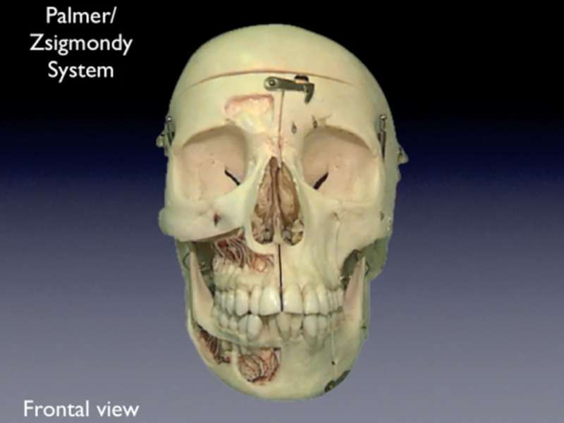
Click on the movie icon above to view the Palmer Zsigmondy shorthand system.
Universal Shorthand System
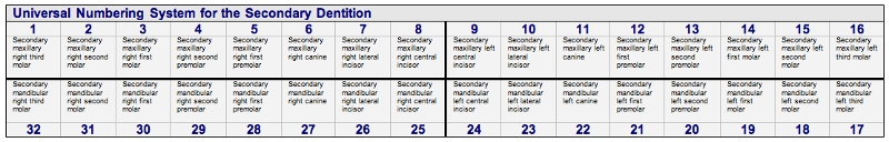
Universal Shorthand for the Secondary Dentition
The Universal (American) shorthand numbering system is the shorthand system used in the United States. Common to other numbering systems, teeth in the secondary (permanent) dentition are numbered in a clockwise fashion from the maxillary right third molar (1) to the mandibular right third molar (32). This system does not use quadrant numbering but uses consecutive numbers from 1 to 32 to number the secondary (permanent) teeth. The mandibular left third molar in the Universal system is 17 and is pronounced "seventeen". The same tooth in the International system is tooth 3.8 and is pronounced tooth "three eight".
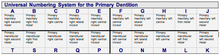
Universal Shorthand Numbering System for the Primary Dentition
The Universal shorthand system for the primary dentition uses capital letters from A-T and follows the same pattern as the secondary (permanent) dentition. The primary maxillary right second molar is represented by the letter A and continues alphabetically in a clockwise fashion to the mandibular right second molar which is represented by the letter T.
Tooth Development
The normal stages of development of each of the primary and secondary teeth are important. The key events for the secondary dentition are shown below. Although there is a range of normal, this knowledge can assist in determining whether your patient's dentition is developing normally or not. Parents will frequently ask about whether their child's dentition is developing normally. In addition, this knowledge is invaluable in determining whether Individual teeth are congenitally missing. Aberrations in the timing of development may also provide insight into systemic problems.
Summary Table of Secondary Tooth Development - Key Timing
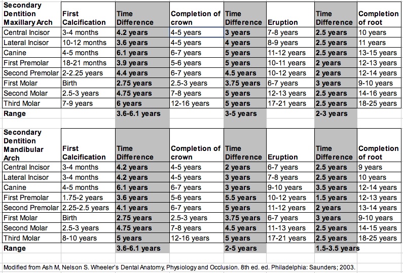
Calcification of all of the teeth in the secondary dentition occurs from birth (secondary maxillary and mandibular first molars) to approximately 10 years of age (maxillary third molar). In this Summary Table of Tooth Development - Key Timing, there is a range of 3.6-6.1 years between the time of initial calcification of the crown until the crown is completed. On average, it takes anywhere from 2-5 years from the time of crown completion until the tooth erupts into the mouth. When teeth erupt, there is approximately 1/2 to 2/3 of the root formed. After a tooth erupts, further root formation occurs. On average, it takes 1.5-3.5 years for the root to completely form after tooth eruption. Variations among the timing of development for each of the secondary teeth are shown in the table above.
Self-Test on Calcification Timing
Self-Test on Eruption Timing
Dental Formula for the Human Dentition
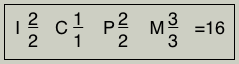
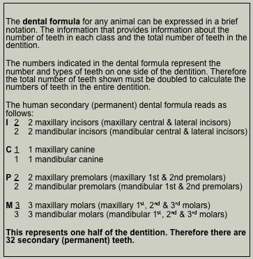
The number and classes of teeth vary among mammals and the dental formula is used to differentiate humans from other mammalian species. Each of the two sets of teeth in the human (primary and secondary) can be expressed by their own dental formula. The formula expresses one half of the dentition and identifies class and the number of those teeth in each class. The total number of teeth in the formula is doubled to express the total number of teeth in the dentition.
The secondary (permanent) human dental formula is
The primary (deciduous) human dental formula is

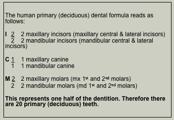
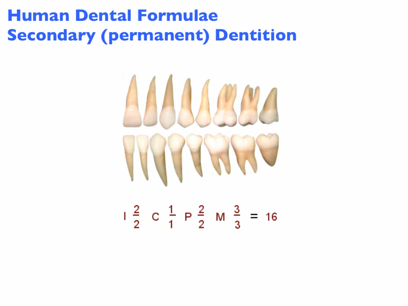
Click on the movie icon above to view the human dental formulae.